INFORMATION
Patient Name: Jim ××
Sex: Male
Age: 61 year-old
Chief Complaints:The Teeth Number 11, 16, and 17 are missing.This treatment aims for the tooth Number 11 implantation.
Clinic Examination
Intro Oral Examination:
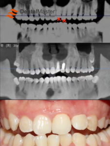
The patient has medium bone quality, with tooth number 11 missing. Bone defects are present on the buccal side of tooth number 11. A DME Φ3.75×13mm implant will be placed in this location, and simultaneous bone grafting will be necessary. Additionally, teeth numbers 16 and 17 are missing. This will be the patient’s first dental implant procedure. The patient has no negative oral habits or previous dental complications; however, they do smoke, which has resulted in some staining and calculus buildup on the teeth. Fortunately, the gums are healthy, with no signs of swelling.
Imaging Examination:
Imaging reveals the absence of tooth number 11 in the anterior region, along with bone defects on the buccal side. The surrounding soft tissues appear to be in good condition.

TREATMENT
Pre-op Planning
Implantation and GBR (Guided Bone Regeneration) Procedure for Tooth Number 11
PROCEDURE
Pre-op Planning
Implantation and GBR Operation in Teeth Number 11
PROCEDURE
1. Cut the gums under local anesthesia.
The bone is medium bone and there are bone defects on the buccal side

2. Use drills to expand the hole gradually.
Implant φ3.75*13mm DME
Drill nourishing holes

3. Fill with 0.25g Bio-Oss bone powder
Cover with T-shape Titanium Mesh

4. Place 1mm Cover Screw

5. Place 2*25 cm colleagen membrane
Make Tension-reduced Suture in tissue flap

Immediate Post-op Radiograph
The X-ray taken immediately after surgery clearly shows the state of the maxillary anterior area after titanium mesh guided bone regeneration (GBR) surgery. In the X-ray, it can be observed that the titanium mesh has been accurately placed in the predetermined position, its structure is complete and stable, and it effectively covers the surgical area. The bone structure around and below the titanium mesh shows good alignment and tightness. At the same time, the soft tissue contours of the surgical area are clear, and no abnormal shadows or displacement are observed, indicating that the surgical operation is delicate and the surrounding tissues are properly protected.


RE-EXAMINATION
To ensure the smooth healing and recovery of the surgical site, the patient needs to return to the hospital for stitch removal after 1 week according to the doctor’s advice. Stitch removal is an important step in postoperative recovery. It marks the initial healing of the surgical wound and lays the foundation for subsequent review and repair work.
After 6 months, the patient needs to return to the hospital for a review again. At this time, the doctor will comprehensively evaluate the bone regeneration and surgical results through X-rays and clinical examinations. If the bone regeneration is good and the surgical site has fully recovered, the patient will receive further cosmetic restoration treatment. Cosmetic restoration aims to improve the appearance and function of the surgical area so that the patient can obtain satisfactory treatment results.

